
See the Diagnosis. Master the Language.
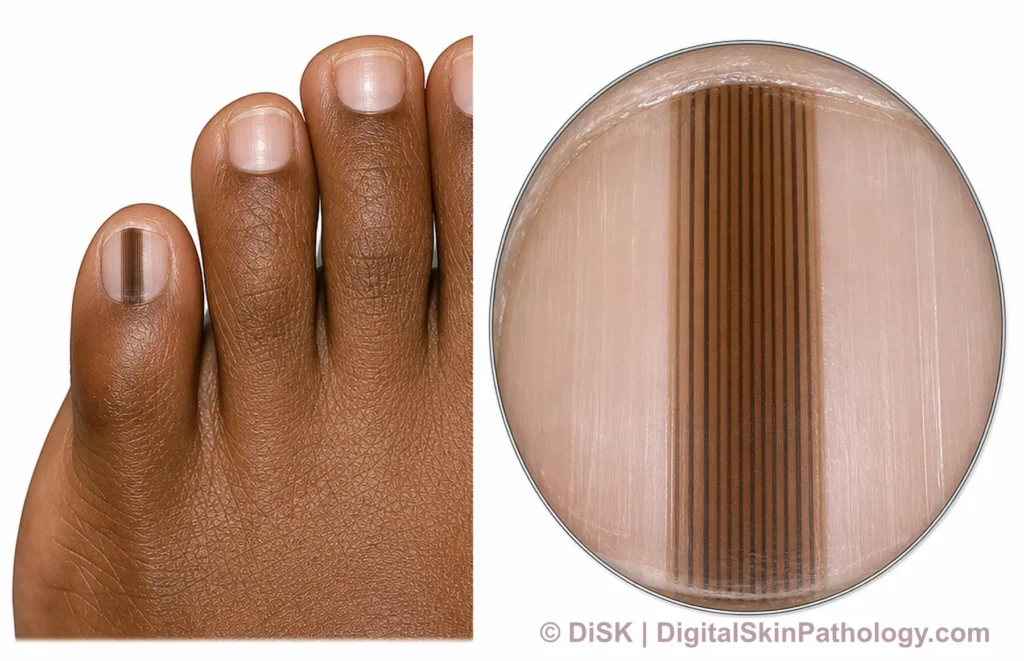
The Challenge: Dermatopathology relies on a specialized lexicon to describe microscopic patterns. For visual learners—a common trait among dermatologists and pathologists—this terminology can sometimes obscure rather than clarify.
Our Method: This growing collection uses precise medical illustration to decode that language. Each plate links a key term directly to its visual signature.
The Pathway: Begin by demystifying terminology through imagery. Then, see how visual features build diagnostic criteria. This foundational knowledge prepares you for confident application in real-world cases.
📚 Explore Our Visual Learning Library
How to Use This Library
- Start with Foundational Terms to learn the basic visual vocabulary
- Move to Specific Diagnoses to see complete entities
- Test yourself with Look-Alikes
- Review with Summary Infographics