
HMB-45 vs. PRAME: How Do They Actually Stack Up?
A question I get from residents almost every week on the fellowship floor: “Do I still need HMB-45, or has PRAME made it obsolete?” The honest answer is neither stain replaces the other — they measure different things, and knowing why changes how you read your slides.
How They Differ
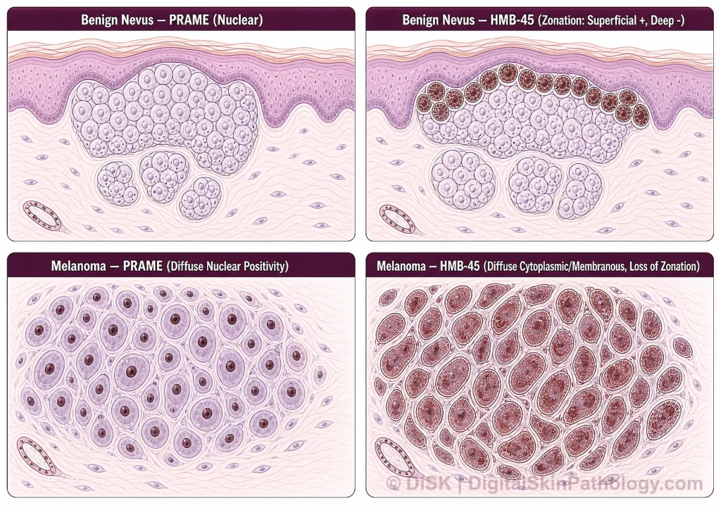
HMB-45 is a cytoplasmic/membranous marker against gp100, a melanosomal glycoprotein. Its major limitation in primary cutaneous melanocytic lesions is a maturation problem: benign nevi normally show strong HMB-45 positivity in the superficial component that fades with descent into the dermis — the so-called maturation pattern. That fading is expected and reassuring. The diagnostic red flag is when a lesion loses this gradient and stains diffusely and strongly throughout its full depth.
PRAME (Preferentially Expressed Antigen in Melanoma) is a nuclear stain, which on its own makes it easier to score than HMB-45’s cytoplasmic pattern — nuclear positivity is binary and harder to confuse with background pigment or histiocytes. PRAME is preferentially expressed in melanoma and largely absent in benign nevi, giving it better specificity for malignancy than HMB-45 alone. It isn’t perfect either: some Spitz nevi, blue nevi, and dysplastic nevi can show patchy PRAME positivity, so a positive result still needs architectural and clinical context, not a standalone yes/no read.
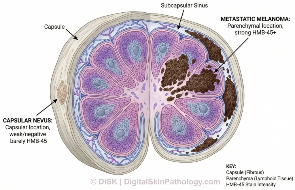
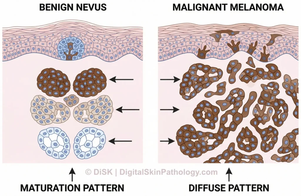
PRAME and HMB-45, Side by Side
The comparison below puts both stains through the same two scenarios — benign nevus and melanoma — so the difference in what each marker is actually measuring becomes visual rather than abstract.
The Practical Takeaway
Neither stain works well in isolation for a genuinely ambiguous lesion. The literature increasingly supports combining PRAME’s nuclear pattern with HMB-45’s depth-dependent staining gradient — benign lesions are rarely diffusely positive for both, while true melanomas much more often are. Used together, they raise diagnostic specificity well beyond either stain alone.
Bottom line: HMB-45 tells you about depth-dependent maturation; PRAME tells you about nuclear expression largely restricted to malignant melanocytes. Neither is a standalone answer key — but together, read in the context of architecture and clinical history, they meaningfully sharpen the call in a genuinely borderline lesion.
Further Reading
For residents who want to go deeper into the primary literature behind this comparison:
- Rasic D, et al. Diagnostic utility of combining PRAME and HMB-45 stains in primary melanocytic tumors. Ann Diagn Pathol. 2023.
- Grillini M, et al. HMB45/PRAME, a Novel Double Staining for the Diagnosis of Melanocytic Neoplasms. Appl Immunohistochem Mol Morphol. 2022.
- PRAME Immunohistochemistry in Thin Melanomas Compared to Melanocytic Nevi. 2024.
Written by Dr. Soheil Dadras